UQsOpticalsMicromanipulationsgroup

Optical Tweezers at the limits

Optical Trapping (OT) is a well-established method for manipulating nanometre to micrometre-scale transparent objects. It uses an optical force known as the gradient force to trap and hold tiny particles in a focused laser beam.

A highly focused laser beam creates an intensity gradient near its focal point sufficiently large to give rise to forces on transparent objects with different refractive indices to their surroundings. The size of particles which they can trap (micro and nano) and the range of forces they exert (piconewto, pN) make them an ideal tool for exploration of a variety of fields such as Biomedical research, Cell Biology, Statistical thermodynamics and colloid science. OT provides one of the most sensitive ways to measure the forces which are present in the microscopic world. Birefringent materials like Vaterite can be used to create tiny micro-rotators which can be used as probes.However the exertion of stronger forces, and the manipulation of larger objects remains challenging. Furthermore, trapping objects in vivo, especially at depth, is difficult because of the scattering and power loss that results from passage through biological tissue.

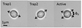
The most common optical trap shape is a focussed Gaussian beam. A Gaussian beam can be used to grab particles, and for non-spherical or birefringent particles, the polarisation of the beam can be used to align the particle. For rotating particles, beams with circular polarisation or optical orbital angular momentum can be used. In order to control the orientation of particles more precily we can explore different shaped traps. Recently we explored the use of annular beam optical tweezers (Lenton et al., 2020) with slowly varying position for the orientation of E coli. Although we found these traps are not very suitable for orientation of E coli in water, they may be useful for orientation or trapping of rod-shaped spores in air or vacuum (in a setup similar to (Pan et al., 2019)).

Example of an optically trapped vaterite.
Artistic impression of otolith in an optical trap.

Recent results include:

(Favre-Bulle et al., 2017) (Favre-Bulle et al., 2015) (Zhang et al., 2017) (Bennett et al., 2013)

2020

  • Orientation of swimming cells with annular beam optical tweezers

    Lenton, I. C. D., Armstrong, D. J., Stilgoe, A. B., Nieminen, T. A., & Rubinsztein-Dunlop, H. (2020). Opt. Commun., 459, 124864. https://doi.org/10.1016/j.optcom.2019.124864'

    Optical tweezers are a versatile tool that can be used to manipulate small particles including both motile and non-motile bacteria and cells. The orientation of a non-spherical particle within a beam depends on the shape of the particle and the shape of the light field. By using multiple beams, sculpted light fields or dynamically changing beams, it is possible to control the orientation of certain particles. In this paper we discuss the orientation of the rod-shaped bacteria Escherichia coli (E. coli) using dynamically shifting annular beam optical tweezers. We begin with examples of different beams used for the orientation of rod-shaped particles. We discuss the differences between orientation of motile and non-motile particles, and explore annular beams and the circumstances when they may be beneficial for manipulation of non-spherical particles or cells. Using simulations we map out the trajectory the E. coli takes. Estimating the trap stiffness along the trajectory gives us an insight into how stable an intermediate rotation is with respect to the desired orientation. Using this method, we predict and experimentally verify the change in the orientation of motile E. coli from vertical to near-horizontal with only one intermediate step. The method is not specific to exploring the orientation of particles and could be easily extended to quantify the stability of an arbitrary particle trajectory.

    @article{Lenton2020Mar,
      author = {Lenton, Isaac C. D. and Armstrong, Declan J. and Stilgoe, Alexander B. and Nieminen, Timo A. and Rubinsztein-Dunlop, Halina},
      title = {{Orientation of swimming cells with annular beam optical tweezers}},
      journal = {Opt. Commun.},
      volume = {459},
      pages = {124864},
      year = {2020},
      month = mar,
      issn = {0030-4018},
      publisher = {North-Holland},
      doi = {10.1016/j.optcom.2019.124864}
    }
    

2019

  • Optical-trapping of particles in air using parabolic reflectors and a hollow laser beam

    Pan, Y.-L., Kalume, A., Lenton, I. C. D., Nieminen, T. A., Stilgoe, A. B., Rubinsztein-Dunlop, H., Beresnev, L. A., Wang, C., & Santarpia, J. L. (2019). Opt. Express, 27(23), 33061–33069. https://doi.org/10.1364/OE.27.033061'

    We present an advanced optical-trapping method that is capable of trapping arbitrary shapes of transparent and absorbing particles in air. Two parabolic reflectors were used to reflect the inner and outer parts of a single hollow laser beam, respectively, to form two counter-propagating conical beams and bring them into a focal point for trapping. This novel design demonstrated high trapping efficiency and strong trapping robustness with a simple optical configuration. Instead of using expensive microscope objectives, the parabolic reflectors can not only achieved large numerical aperture (N.A.) focusing, but were also able to focus the beam far away from optical surfaces to minimize optics contamination. This design also offered a large free space for flexible integration with other measuring techniques, such as optical-trapping Raman spectroscopy, for on-line single particle characterization.

    @article{Pan2019Nov,
      author = {Pan, Yong-Le and Kalume, Aimable and Lenton, Isaac C. D. and Nieminen, Timo A. and Stilgoe, Alex B. and Rubinsztein-Dunlop, Halina and Beresnev, Leonid A. and Wang, Chuji and Santarpia, Joshua L.},
      title = {{Optical-trapping of particles in air using parabolic reflectors and a hollow laser beam}},
      journal = {Opt. Express},
      volume = {27},
      number = {23},
      pages = {33061--33069},
      year = {2019},
      month = nov,
      issn = {1094-4087},
      publisher = {Optical Society of America},
      doi = {10.1364/OE.27.033061}
    }
    

2017

  • Optical trapping of otoliths drives vestibular behaviours in larval zebrafish

    Favre-Bulle, I. A., Stilgoe, A. B., Rubinsztein-Dunlop, H., & Scott, E. K. (2017). Nature Communications, 8(1). https://doi.org/10.1038/s41467-017-00713-2'

    The vestibular system, which detects gravity and motion, is crucial to survival, but the neural circuits processing vestibular information remain incompletely characterised. In part, this is because the movement needed to stimulate the vestibular system hampers traditional neuroscientific methods. Optical trapping uses focussed light to apply forces to targeted objects, typically ranging from nanometres to a few microns across. In principle, optical trapping of the otoliths (ear stones) could produce fictive vestibular stimuli in a stationary animal. Here we use optical trapping in vivo to manipulate 55-micron otoliths in larval zebrafish. Medial and lateral forces on the otoliths result in complementary corrective tail movements, and lateral forces on either otolith are sufficient to cause a rolling correction in both eyes. This confirms that optical trapping is sufficiently powerful and precise to move large objects in vivo, and sets the stage for the functional mapping of the resulting vestibular processing.

    @article{Favre-Bulle2017,
      author = {Favre-Bulle, Itia A. and Stilgoe, Alexander B. and Rubinsztein-Dunlop, Halina and Scott, Ethan K.},
      doi = {10.1038/s41467-017-00713-2},
      issn = {20411723},
      journal = {Nature Communications},
      mendeley-groups = {Optical Tweezers/UQOMG},
      month = dec,
      number = {1},
      publisher = {Nature Publishing Group},
      title = {{Optical trapping of otoliths drives vestibular behaviours in larval zebrafish}},
      volume = {8},
      year = {2017}
    }
    
  • Ultrasensitive rotating photonic probes for complex biological systems

    Zhang, S., Gibson, L. J., Stilgoe, A. B., Favre-Bulle, I. A., Nieminen, T. A., & Rubinsztein-Dunlop, H. (2017). Optica, 4(9), 1103. https://doi.org/10.1364/optica.4.001103'

    \textcopyright 2017 Optical Society of America. We use rotational photonic tweezers to access local viscoelastic properties of complex fluids over a wide frequency range. This is done by monitoring both passive rotational Brownian motion and also actively driven transient rotation between two angular trapping states of a birefringent microsphere. These enable measurement of high- and low-frequency properties, respectively. Complex fluids arise frequently in microscopic biological systems, typically with length scales at the cellular level. Thus, high spatial resolution as provided by rotational photonic tweezers is important. We measure the properties of tear film on a contact lens and demonstrate variations in these properties between two subjects over time. We also show excellent agreement between our theoretical model and experimental results. We believe that this is the first time that active microrheology using rotating tweezers has been used for biologically relevant questions. Our method demonstrates potential for future applications to determine the spatial-temporal properties of biologically relevant and complex fluids that are only available in very small volumes.

    @article{Zhang2017,
      author = {Zhang, Shu and Gibson, Lachlan J. and Stilgoe, Alexander B. and Favre-Bulle, Itia A. and Nieminen, Timo A. and Rubinsztein-Dunlop, Halina},
      doi = {10.1364/optica.4.001103},
      issn = {2334-2536},
      journal = {Optica},
      mendeley-groups = {Optical Tweezers/UQOMG},
      month = sep,
      number = {9},
      pages = {1103},
      publisher = {The Optical Society},
      title = {{Ultrasensitive rotating photonic probes for complex biological systems}},
      volume = {4},
      year = {2017}
    }
    

2015

  • Scattering of sculpted light in intact brain tissue, with implications for optogenetics

    Favre-Bulle, I. A., Preece, D., Nieminen, T. A., Heap, L. A., Scott, E. K., & Rubinsztein-Dunlop, H. (2015). Scientific Reports, 5. https://doi.org/10.1038/srep11501'

    Optogenetics uses light to control and observe the activity of neurons, often using a focused laser beam. As brain tissue is a scattering medium, beams are distorted and spread with propagation through neural tissue, and the beam’s degradation has important implications in optogenetic experiments. To address this, we present an analysis of scattering and loss of intensity of focused laser beams at different depths within the brains of zebrafish larvae. Our experimental set-up uses a 488 €‰nm laser and a spatial light modulator to focus a diffraction-limited spot of light within the brain. We use a combination of experimental measurements of back-scattered light in live larvae and computational modelling of the scattering to determine the spatial distribution of light. Modelling is performed using the Monte Carlo method, supported by generalised Lorenz-Mie theory in the single-scattering approximation. Scattering in areas rich in cell bodies is compared to that of regions of neuropil to identify the distinct and dramatic contributions that cell nuclei make to scattering. We demonstrate the feasibility of illuminating individual neurons, even in nucleus-rich areas, at depths beyond 100\mum using a spatial light modulator in combination with a standard laser and microscope optics.

    @article{Favre-Bulle2015,
      author = {Favre-Bulle, Itia A. and Preece, Daryl and Nieminen, Timo A. and Heap, Lucy A. and Scott, Ethan K. and Rubinsztein-Dunlop, Halina},
      doi = {10.1038/srep11501},
      issn = {20452322},
      journal = {Scientific Reports},
      mendeley-groups = {Optical Tweezers/UQOMG},
      month = jun,
      publisher = {Nature Publishing Group},
      title = {{Scattering of sculpted light in intact brain tissue, with implications for optogenetics}},
      volume = {5},
      year = {2015}
    }
    

2013

  • Spatially-resolved rotational microrheology with an optically-trapped sphere

    Bennett, J. S., Gibson, L. J., Kelly, R. M., Brousse, E., Baudisch, B., Preece, D., Nieminen, T. A., Nicholson, T., Heckenberg, N. R., & Rubinsztein-Dunlop, H. (2013). Scientific Reports, 3. https://doi.org/10.1038/srep01759'

    We have developed a microrheometer, based on optical tweezers, in which hydrodynamic coupling between the probe and fluid boundaries is dramatically reduced relative to existing microrheometers. Rotational Brownian motion of a birefringent microsphere within an angular optical trap is observed by measuring the polarisation shifts of transmitted light. Data gathered in this manner, in the strongly viscoelastic fluid Celluvisc, quantitatively agree with the results of conventional (bulk) rheometry. Our technique will significantly reduce the smallest sample volumes which may be reliably probed, further extending the study of rare, difficult to obtain or highly nonhomogeneous fluids.

    @article{Bennett2013,
      author = {Bennett, James S. and Gibson, Lachlan J. and Kelly, Rory M. and Brousse, Emmanuel and Baudisch, Bastian and Preece, Daryl and Nieminen, Timo A. and Nicholson, Timothy and Heckenberg, Norman R. and Rubinsztein-Dunlop, Halina},
      doi = {10.1038/srep01759},
      issn = {20452322},
      journal = {Scientific Reports},
      mendeley-groups = {Optical Tweezers/UQOMG},
      title = {{Spatially-resolved rotational microrheology with an optically-trapped sphere}},
      volume = {3},
      year = {2013}
    }